
Fig. 1
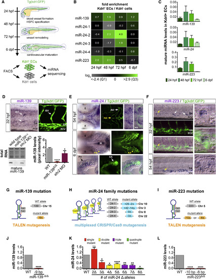
Generation of Endothelial miRNA Mutants in Zebrafish (A) Experimental procedure to identify miRNAs expressed in fluorescence-activated cell (FAC)-sorted Kdrl:GFP+ endothelial cells and Kdrl:GFP− non-endothelial cells during the four major stages of zebrafish vascular development. Dashed boxes outline the regions examined for cardiovascular phenotypes in endothelial miRNA mutant embryos (see Figure S2C). (B) Heatmap depicts miRNA reads per million in Kdrl:GFP+ endothelial relative to non-endothelial cells for two biological replicates. Color scale ranges from the first quartile (Q1) to the third quartile (Q3) fold-enrichment values for all 46 endothelial miRNAs identified (see Figure S1A). (C) Average mature miRNA levels relative to U6 small nuclear RNA (snRNA) expression as determined by qRT-PCR in FAC-sorted Kdrl:GFP+ endothelial cells at the indicated developmental stages. (D) Top: Lateral trunk view (20×) of wild-type embryos showing whole-mount in situ hybridization (WISH) for mature miR-139, labeling cells within the ISV position. Confocal image (25×) shows the relative position of ISVs in the lateral trunk. Bottom: northern blot and respective quantification showing mature miR-139 expression relative to total RNA in three biological replicates of 32-hpf embryos treated as indicated. Consistent with miR-139 expression in ISVs, mature miR-139 levels were diminished in etv2 morphant embryos, which lack these trunk vessels (Pham et al., 2007). (E) Mature miR-24 localization in the ventral head of wild-type embryos in relation to Kdrl:GFP+ vasculature at the indicated developmental stages (20×). By 6 dpf, miR-24 remained in vascular cells but was excluded from cartilaginous and bone structures. Arrows point to anatomic landmarks. Yellow arrows point to the region captured in zoomed-in images (40×). (F) Mature miR-223 localization in the lateral Kdrl:GFP+ trunk vasculature of wild-type embryos at the indicated stages (20×). At 54 hpf, miR-223 is expressed in cells within the caudal hematopoietic tissue located between the DA and CV. Arrows show examples of miR-223+ cells. Yellow arrows point to the region captured in magnified images. (G–I) Schematic representation of the genome-editing strategies employed to mutagenize endothelial miRNAs. TALENs and a multiplexed CRISPR/Cas9 system were targeted to miRNA precursor genomic sequences to prevent mature miRNA formation. Gray boxes represent the wild-type allele. Colored boxes reveal the nature of the mutant allele. See also Figure S2A. (J–L) qRT-PCR showing average mature miRNA expression normalized to U6 snRNA levels in miRNA mutant embryos (J), embryo heads (K), or adult fins (L) relative to wild-type. For miR-24 mutants, genotypes were categorized as a single mutant when two (Δ2, miR-24-4Δ/Δ) and three (Δ3, e.g., miR-24-1+/Δ 4Δ/Δ) miR-24 alleles were mutated, up to a quadruple mutant that lacked all eight miR-24 alleles (Δ8, miR-24-1Δ/Δ 2Δ/Δ 3Δ/Δ 4Δ/Δ). See also Figure S2B. Bar plots represent mean ± SEM and significance calculations are relative to wild-type embryos. n.s., not significant (p > 0.05); ∗p ≤ 0.05, ∗∗p ≤ 0.01, ∗∗∗∗p ≤ 0.0001, two-tailed Student's t test. qRT-PCR data represent two to five biological replicates. Ten to 20 embryos from at least two different clutches were examined by WISH. AA, aortic arches; DA, dorsal aorta; CV caudal vein; E, eye; EC, endothelial cell; H, heart; HA, hypobranchial artery; ISV, intersegmental vessel; PA, pharyngeal arch; PCV, posterior cardinal vein. |
| Genes: | |
|---|---|
| Fish: | |
| Anatomical Terms: | |
| Stage Range: | Prim-15 to Day 6 |
Reprinted from Developmental Cell, 40, Kasper, D.M., Moro, A., Ristori, E., Narayanan, A., Hill-Teran, G., Fleming, E., Moreno-Mateos, M., Vejnar, C.E., Zhang, J., Lee, D., Gu, M., Gerstein, M., Giraldez, A., Nicoli, S., MicroRNAs Establish Uniform Traits during the Architecture of Vertebrate Embryos, 552-565.e5, Copyright (2017) with permission from Elsevier. Full text @ Dev. Cell