
Fig. S4
- ID
- ZDB-FIG-170505-43
- Publication
- Armstrong et al., 2017 - Shh promotes direct interactions between epidermal cells and osteoblast progenitors to shape regenerated zebrafish bone
- Other Figures
- All Figure Page
- Back to All Figure Page
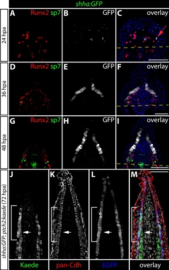
Spatial and temporal analysis of shha:GFP expression during fin regeneration. (A-I) Caudal fin sections from shha:GFP fish at 24 (A-C), 36 (D-F), and 48 (G-I) hpa immunostained with antibodies against Runx2 (red), sp7 (green) and GFP (white) and imaged by confocal microscopy. A red arrow indicates a GFP+ basal epidermal cell at 24 hpa. By 36 hpa, nearly every basal epidermal cell is GFP+. Amputation planes are marked with a dashed yellow line. Hoechst-stained nuclei (blue) are shown in the overlay panels. (J-M) An immunostained fin section from a 72 hpa shha:GFP;ptch2:Kaede fish showing pan-cadherin (red), Kaede (green), and GFP (blue). The white arrow indicates the distal extent of Kaede-expressing osteoblasts. The white bracket shows basal epidermis with ptch2-driven Kaede protein. Hoechst-stained nuclei are grey in the overlay panel. Scale bars: 50 μm. |