
Fig. S1
- ID
- ZDB-FIG-170505-40
- Publication
- Armstrong et al., 2017 - Shh promotes direct interactions between epidermal cells and osteoblast progenitors to shape regenerated zebrafish bone
- Other Figures
- All Figure Page
- Back to All Figure Page
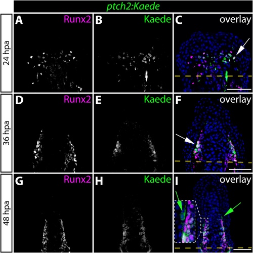
Hedgehog/Smoothened signaling initiates in osteoblast progenitors before expanding to neighboring basal epidermal cells during the course of fin regeneration. (A-I) Immunostaining showing Runx2 (magenta) and Kaede (green) protein in fin sections prepared from ptch2:Kaede fish at 24 (A-C), 36 (D-F), and 48 (G-I) hours post-amputation (hpa). Ptch2:Kaede is first expressed in Runx2+ pObs at 24 hpa and is excluded from all other cell types. By 36 hpa, nearly all Runx2+ cells co-express Kaede. White arrows indicate Runx2+/Kaede+ pObs. By 48 hpa, ptch2:Kaede is also expressed in the basal epidermis adjacent to Kaede+/Runx2+ pObs. Green arrows point to Kaede+ basal epidermal cells. For panel I, the region bound by a dashed white box is shown in higher magnification in the inset panel. Amputation planes are indicated with a dashed yellow line. Hoechst-stained nuclei are blue in all overlay images. Scale bars: 50 μm. |