
Fig. 1
- ID
- ZDB-FIG-170308-14
- Publication
- Madelaine et al., 2017 - The hypothalamic NPVF circuit modulates ventral raphe activity during nociception
- Other Figures
- All Figure Page
- Back to All Figure Page
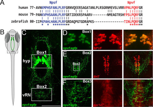
NPVF circuit interacts with the raphe nucleus. (A) Alignment of human, mouse and zebrafish NPVF precursors. Multiple sequence alignments show strong conservation in the sequence of putative NPSF (blue) and NPVF (red) neuropeptides. Stars indicate sequence identity across the alignment. (B) Schematic of larval zebrafish, with location of NPVF neurons (hyp.: hypothalamus; vRN: ventral raphe nucleus). (C) Confocal Z-projection of Tg(npvf:EGFP) larval fish at 5 dpf. Hypothalamus and vental raphe nucleus are highlighted in Box 1 and 2 respectively. (D) Confocal overlay of EGFP+ neurons and NPVF peptide in the hypothalamus at 5 dpf. (E) eGFP+ axons and 5HT antibody stain in the raphe nucleus at 5 dpf. (F) Neurons in the vRN are positive for in situ hybridization for gpr147 at 5 dpf. Scale bars: C = 100 μm, D, E and F = 10 μm. |
| Genes: | |
|---|---|
| Antibody: | |
| Fish: | |
| Anatomical Terms: | |
| Stage: | Day 5 |