
Fig. 1
- ID
- ZDB-FIG-170306-2
- Publication
- Gays et al., 2017 - An exclusive cellular and molecular network governs intestinal smooth muscle cell differentiation in vertebrates
- Other Figures
- All Figure Page
- Back to All Figure Page
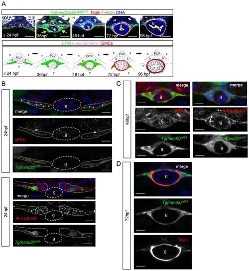
LPM gives rise to iSMCs in zebrafish embryos. (A) Time-course analyses of Tg(hand2:EGFP)pd24 and iSMC marker expression (Tagln) during intestinal development. Tg(hand2:EGFP)pd24 embryos were fixed at different time points from 24 hpf until 96 hpf. Upper panel: confocal transverse sections of the posterior gut region between the somites 7 and 13 of Tg(hand2:EGFP)pd24 embryos stained with phalloidin (gray) and Tagln (red) (single channels are shown in Fig. S1A). The dashed yellow lines highlight LPM/hand2+ cells, whereas the dashed white lines highlight the enteric endoderm (g). Migration of the LPM is indicated by arrows. Asterisks indicate single-cell nuclei. ISMC differentiation is visible during intestinal development by expression of Tagln; blue indicates nuclei; g, gut. Scale bar: 30 μm. Bottom panel: schematic representation of LPM/hand2+ conversion to iSMCs in the gut region of developing zebrafish embryos. Green, LPM; pink, endoderm; red, iSMCs; p, pronephros; s, somite; PCV, posterior cardinal vein; y, yolk. (B) Analyses of Tg(hand2:EGFP)pd24 and polarity and mesenchymal markers during LPM development at 24 hpf. Confocal transverse sections of the posterior gut region between the somites 7 and 13 of Tg(hand2:EGFP)pd24 embryos stained with aPKC or N-cadherin. Nuclei are in blue; g, gut. Scale bars: 30 μm. Asterisks indicate single-cell nuclei while the dashed yellow lines highlight LPM/hand2+ cells. (C) Analyses of Tg(hand2:EGFP)pd24 and polarity and mesenchymal markers during LPM development at 48 hpf. Confocal transverse sections of the posterior gut region between the somites 7 and 13 of Tg(hand2:EGFP)pd24 embryos stained with aPKC (left, red) or N-cadherin (right, red). Blue indicates nuclei; g, gut. Scale bars: 30 μm. (D) Analyses of Tg(hand2:EGFP)pd24 and iSMC marker expression (Tagln) at 72 hpf. Confocal transverse sections of the posterior gut region between the somites 7 and 13 of Tg(hand2:EGFP)pd24 embryos stained with Tagln (red) show that all differentiated iSMC are also Tg(hand2:EGFP)pd24 positive. These observations suggest that posterior LPM expression of hand2 does not demarcate the entire LPM, but rather is confined to the presumptive iSMC progenitors from its expression onset after LPM formation. Nuclei are in blue; g, gut. Scale bars: 30 μm. |
| Gene: | |
|---|---|
| Antibody: | |
| Fish: | |
| Anatomical Terms: | |
| Stage Range: | Prim-5 to Day 4 |