
Fig. 3
- ID
- ZDB-FIG-170222-59
- Publication
- Reade et al., 2017 - TAEL: A zebrafish-optimized optogenetic gene expression system with fine spatial and temporal control
- Other Figures
- All Figure Page
- Back to All Figure Page
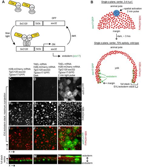
Conversion of presumptive EVL and ectoderm to endoderm via TAEL-dependent Sox32 expression. (A) Schematic depicting experimental set-up for light-induced endoderm induction. Tg(cryaa:Venus;C120:sox32); Tg(sox17:GFP) embryos were injected with TAEL mRNA. Ectoderm progenitors at the animal pole were illuminated with blue light, leading to misexpression of sox32 and adoption of endoderm fate (expression of sox17:GFP). (B) Top panel depicts an embryo at 3-4 hpf with nuclei labeled by H2B-mCherry (red). Activating light was restricted to the animal pole (blue circle, approximately 116 μm in diameter). Bottom panel depicts a transverse cross section of an embryo at 70-80% epiboly. Endodermal cells are labeled by Tg(sox17:GFP) (green) and all nuclei are labeled with H2B-mCherry (red). Brackets labeled ‘EVL/ectoderm stack’ and ‘Full stack’ indicate approximate positions of images shown in C-K and L-N, respectively. (C-K) Maximum z-projections of EVL and ectoderm layers only. Embryos lacking TAEL (C) or expressing TAEL but kept in the dark (D) show no GFP-positive cells in these layers. However, activated embryos (E) exhibit numerous GFP-positive cells within the EVL and ectoderm domain. (L-N) Re-sliced x-z views of the entire acquired stack (EVL/ectoderm towards the top, endoderm toward the bottom). In embryos lacking TAEL (L) or expressing TAEL but kept in the dark (M) GFP-positive cells are restricted to a single endodermal layer adjacent to the yolk, whereas in activated embryos (N), GFP-positive cells are located in multiple cell layers, especially in the outer EVL and ectoderm domains. Uninjected, 17% with ectopic endoderm, n=17; dark, 33% with ectopic endoderm, n=9; light activated, 65% with ectopic endoderm, n=17. |