
Fig. 3
- ID
- ZDB-FIG-170203-13
- Publication
- Miao et al., 2017 - Translation repression by maternal RNA binding protein zar1 is essential for early oogenesis in zebrafish
- Other Figures
- All Figure Page
- Back to All Figure Page
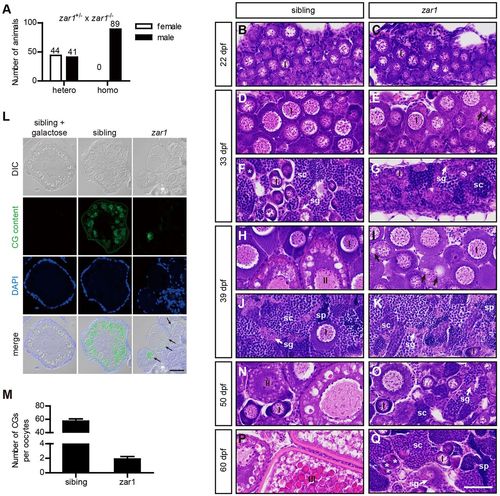
Zar1 deficiency causing all-male phenotype is due to female-to-male sex reversal. (A) Analysis of genders of zar1 homozygotes (homo) and heterozygous siblings (hetero). zar1+/− heterozygous females were crossed with zar1−/− homozygous males and genders of their progenies were analyzed. (B-K,N-Q) Gonad development of zar1 homozygotes and control siblings analyzed by H&E staining. At 22 dpf, zebrafish gonads are undifferentiated. Gonads of zar1 homozygotes and control siblings are indistinguishable histologically (B,C). At 33 dpf, WT gonads differentiate into immature ovaries and immature testes. Minor developmental abnormalities are observed in zar1 homozygotes. Oocytes in zar1 homozygotes are similar to those in WT in size and morphology, but aberrant vesicles (arrow indicated) are observed in ooplasm of a few zar1 homozygous oocytes (D,E). Testis development in zar1 homozygotes is normal compared with that in control siblings (F,G). At 39 dpf, ovarian developmental abnormality in zar1 homozygotes becomes more pronounced (H,I), while testis development in zar1 homozygotes is similar to the controls (J,K). Immature ovaries in control siblings contain stage I and stage II oocytes (H) while oocytes in zar1 homozygotes resemble stage I oocytes, indicating oogenesis arrest in zar1 homozygotes. In addition, aberrant vesicles (arrow) in zar1 homozygotes increase significantly in size and number. (L) MPA (Maclura pomifera agglutinin) staining of ovary sections. Juveniles at 37-40 dpf fixed with 4% PFA were embedded in paraffin and sections stained with MPA; 0.5 M D-galactose inhibited MPA staining. MPA specifically stains CGs in control siblings. The aberrant vesicles in zar1 mutant ovaries are MPA positive (arrows). (M) Comparison of CG numbers (mean±s.e.m.) between zar1 mutants (n=11) and siblings (n=11). At 50 dpf, stage II oocytes are observed in heterozygous and wild-type ovaries (N), but large numbers of oocytes underwent atresia in zar1 homozygotes, and spermatogonia and spermatocytes appear among oocytes, indicating transitional ovaries (ovotestis) (O). At 60 dpf, stage III oocytes were seen in the control ovaries (P); in contrast, very few oocytes remain in ovotestis of zar1 mutants and spermatogenesis has progessed further (Q). At 22 dpf, six juveniles were analyzed for each genotype. At 33 dpf, 39 dpf, 50 dpf and 60 dpf, 20 juveniles per stage were analyzed for each genotype. Stars indicate degenerating oocytes. sg, spermatogonia; sc, spermatocytes; sp, sperm. Scale bar: 40 μm. |
| Fish: | |
|---|---|
| Observed In: | |
| Stage Range: | Days 30-44 to Adult |