
Fig. 4
- ID
- ZDB-FIG-161219-31
- Publication
- Bremer et al., 2016 - Myosin phosphatase Fine-tunes Zebrafish Motoneuron Position during Axonogenesis
- Other Figures
- All Figure Page
- Back to All Figure Page
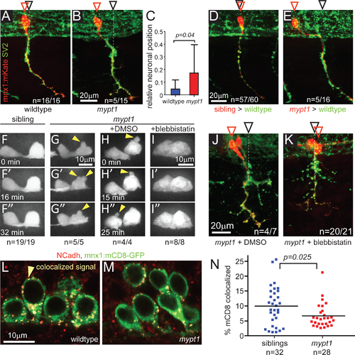
mypt1 is required cell-autonomously for motoneuronal positioning. (A-C) SV2 staining (all axons, green) combined with stochastically labeled CaP motor neurons using mnx1:mKate (single cell labeling, red) to determine relative CaP soma positions in 26 hpf wildtype (A) and mypt1 mutant embryos (B). Red arrowheads indicate the position of CaP cell bodies, black arrowheads the position of the axonal exit point. In contrast to wildtype, 33% of mutant CaP cell bodies were shifted rostrally (p = 0.0177, Fisher exact; for detail on quantification, see Material and Methods). Bar graph of relative neuronal position in wildtype and mypt1 mutants (C). Following transplantation, wildtype-derived CaP motoneurons (labeled with rhodamine-dextran, red) exhibited normal motoneuron positioning in 26–27 hpf wildtype embryos (D). In contrast, mypt1 mutant derived CaP motoneuron when transplated into wildtype embryos frequently failed to adjust their position (E; p = 0.0087, Fisher exact). (F-I) Time-lapse analysis of motoneuron membrane dynamics in 18–19 hpf transgenic mnx1:mKate-mnx1:mCD8-mKate embryos, expressing mKate in the cytoplasm and on cell membranes of motoneurons. Motoneurons in mypt1 mutants displayed membrane blebbing (yellow arrowheads in G, G', G"; n = 5; p = 0.0001, Fisher exact), not observed in wildtype siblings (n = 19, F, F', F"). Treatment with the myosin II inhibitor blebbistatin but not with DMSO (H, H', H") completely abolished membrane blebbing in mypt1 mutants (I, I', I"; n = 8,). (J, K) SV2 staining (all axons, green) combined with stochastically labeled CaP motor neurons using mnx1:mKate (single cell labeling, red) to determine relative CaP soma positions in 26 hpf mypt1 mutant embryos treated with DMSO for control (J) or blebbistatin (K). Blebbistatin treatment significantly reduced rostral mispositioning of mypt1 mutant CaP motoneurons (p = 0.0077, Fisher exact). (L-N) N-Cadherin (NCadh, red) staining of fixed embryos carrying the mnx1:mCD8-GFP transgene (green) which labels motoneuronal membranes. Single plane of confocal images with pseudocolored colocalizing pixels generated by Imaris software in a sibling (L) and a mypt1 mutant (M). Percentage of the green volume (mCD8+) which is colocalized is determined to quantify the fraction of NCadh+ cell membrane (N). |
| Genes: | |
|---|---|
| Antibodies: | |
| Fish: | |
| Condition: | |
| Anatomical Terms: | |
| Stage Range: | 14-19 somites to Prim-5 |
| Fish: | |
|---|---|
| Condition: | |
| Observed In: | |
| Stage Range: | 14-19 somites to Prim-5 |