
Fig. 2
- ID
- ZDB-FIG-161219-29
- Publication
- Bremer et al., 2016 - Myosin phosphatase Fine-tunes Zebrafish Motoneuron Position during Axonogenesis
- Other Figures
- All Figure Page
- Back to All Figure Page
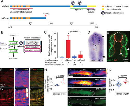
A non-sense mutation in mypt1 causes axon guidance errors. (A) Schematics outlining the domains of the wildtype (top) and the truncated mypt1p82emcf protein (bottom). (B) Phosphorylation-dependent regulation of myosin II: Myosin light chain kinase (MLCK) and Rho kinase (ROCK) increase myosin II phosphorylation and thereby enhance myosin II contractility. Conversely, Myosin phosphatase composed of Mypt1, catalytic and regulatory subunits decreases myosin II phosphorylation and thus causes myosin II relaxation. (C) Injection of 250pg wildtype mypt1 mRNA into one-cell stage wildtype (blue bars) and p82emcf mutant embryos (red bars) significantly reduced motor axon guidance errors in mypt1 mutant embryos as assayed at 26 hpf using SV2 staining (p<0.001; two-tailed t-test). (D) In 25 hpf embryos mypt1 mRNA is readily detectable in the spinal cord (dotted line) and in the myotomes (arrowheads). (E) 3D projection image of a cross section through the trunk of a 26 hpf embryo reveals p-MLC expression in slow-twitch muscle cells (green, arrow head) and in fast-twitch muscle fibers (arrow), while in the spinal cord (dashed circle) p-MLC expression levels are below detection limit. Motor axons are stained with SV2 (in red). (F-H) Co-staining of MHC and p-MLC in siblings (F) and mypt1 mutants (G). Compared to MHC levels (F, G), p-MLC levels are increased in mypt1 mutants (G’, G”) when compared to wildtype (F’, F”; quantified in H). (I-K) FRET analysis using the SECFP donor (I pre, I’ post bleaching) and the YPet acceptor (J pre, J’ post bleaching) reveals increased MLC phosphorylation, quantified in K. Note that non-bleached areas (outside of the region of interest, ROI) remained unchanged. |
| Gene: | |
|---|---|
| Antibody: | |
| Fish: | |
| Anatomical Terms: | |
| Stage: | Prim-5 |
| Fish: | |
|---|---|
| Observed In: | |
| Stage: | Prim-5 |