
Fig. 7
- ID
- ZDB-FIG-161129-14
- Publication
- Marro et al., 2016 - Collagen XII Contributes to Epicardial and Connective Tissues in the Zebrafish Heart during Ontogenesis and Regeneration
- Other Figures
- All Figure Page
- Back to All Figure Page
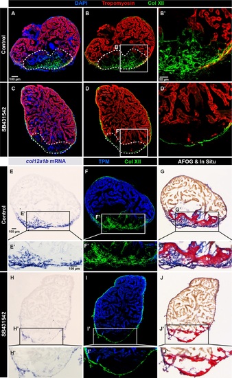
Collagen XII deposition in the post-cryolesion tissue requires TGF-β signaling. (A, B) Immunofluorescence staining shows a normal pattern of Col XII (green) in the Tropomyosin (red)-negative area in control heart. N = 5. (C, D) The treatment with the inhibitor of the TGF-β receptors, SB431542, suppresses the Col XII deposition (green) in the post-cryolesion area, without affecting the pre-existing epicardial fibrils. N = 7. (E-J) In -situ hybridization, double immunostaining and AFOG coloration of control (E-G) and SB431542-treated (H-J) hearts. (E, F) In -situ hybridization and fluorescent staining reveals that col12a1b transcripts (blue) match the distribution of Col XII protein. (F) Col XII fibrils (green) are present in the post-cryolesion area that is recognized by the absence of Tropomyosin (blue). (G) AFOG staining highlights the intact muscle (orange), fibrin-like matrix (red) and collagen (blue) that overlaps with the in -situ staining. (H) The inhibition of TGF-β signaling prevents upregulation of col12a1b expression in the post-cryoinjury area, without affecting its epicardial expression. (I) The cryoinjured zone is devoid of Col XII. (J) AFOG coloration reveals the absence of collagenous matrix (blue) in the post-infarcted zone. N = 6. |