
Fig. 2
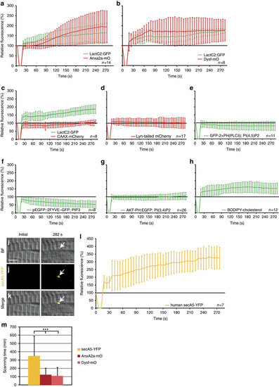
Phosphatidylserine (PS) is sorted to the repair patch. (a-h) Real-time analysis of enrichment of PS (LactC2:GFP (a-c)), PI(4,5)P2 (GFP-2 × PH(PLC´) (e)), PIP3 (pEGFP::2FYVE–GFP (f)), PI(3,4)P2 (AKT-PH:EGFP (g)) and cholesterol (BODIPY-cholesterol (h)), relative to AnxA2a-mO (a), Dysf–mO (b) and membrane markers CAAX-mCherry (c), Lyn-tailed mCherry (d). The reporter fluorescence is expressed as percentage (mean±s.d.) relative to the level before injury. (i-m) PS is presented on the extracellular side of the repair patch. (i–l) Extracellularly supplied secA5-YFP was enriched at membrane on lesioning (i–k, arrows). secA5-YFP expressing myofibers are outside of the field of view. (l) Kinetics of secA5-YFP at the repair patch. (m) Macrophage scanning-time (+/ s.d.) of myofibers expressing secreted secA5-YFP (yellow, n=19), AnxA2a-mO (dark red, n=22) or Dysf–mO (light red, n=23; Student t-test P<0.001) Scale bar, 4 µm. |