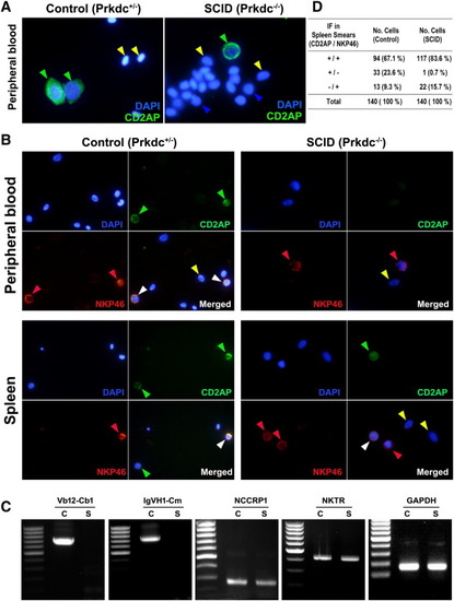
IF and RT-PCR analyses for activated lymphocytes. (A) IF detected by CD2AP marker. The CD2AP-reactive lymphocytes are present in the peripheral blood from both SCID and control zebrafish, suggesting that the cells are either activated T lymphocytes or NK cells. DAPI staining was used to detect the blood cells. (B) Double IF detected by CD2AP (Cy2) and NKP46 (Cy3) markers. The NKP46 is a specific marker for NK cells, whereas the CD2AP marker is reactive to both T lymphocytes and NK cells. (C) RT-PCR analysis. Glyceraldehyde 3-phosphate dehydrogenase gene detection was used as control. Nonspecific cytotoxic cell receptor protein-1 and NK specific protein (NK cell triggering receptor) gene detections indicate NK cell. TCR V(D)JC (Vb12-Cb1) and immunoglobulin (IgVH1-Cm) gene transcript detections reveal V(D)J recombination and heavy-chain rearrangement. (D) Counted numbers of cells depending on CD2AP or NKP46 positivity. Green arrowhead, positive for CD2AP; red arrowhead, positive for NKP46; white arrowhead, positive for both CD2AP and NKP46; blue arrowhead, lymphocytes negative for both CD2AP and NKP46; yellow arrowhead; red blood cells.
|
