
FIGURE
Fig. 2
- ID
- ZDB-FIG-160928-21
- Publication
- Mercatali et al., 2016 - Development of a Patient-Derived Xenograft (PDX) of Breast Cancer Bone Metastasis in a Zebrafish Model
- Other Figures
- All Figure Page
- Back to All Figure Page
Fig. 2
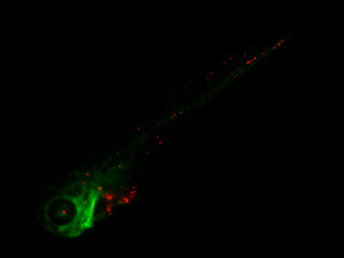
Representative image of MDA-MB-231 cell line five days after injection into the duct of Couvier of 2 day post fertilization (dpf), Tg(fli1:GFP) ZF embryo. MDA-MB-231, labeled in red, were monitored on a daily basis for the duration of the experiment (five days) and showed progressive and extensive dissemination throughout the developing embryo (25× magnification). |
Expression Data
| Gene: | |
|---|---|
| Fish: | |
| Condition: | |
| Anatomical Term: | |
| Stage: | Long-pec |
Expression Detail
Antibody Labeling
Phenotype Data
Phenotype Detail
Acknowledgments
This image is the copyrighted work of the attributed author or publisher, and
ZFIN has permission only to display this image to its users.
Additional permissions should be obtained from the applicable author or publisher of the image.
Full text @ Int. J. Mol. Sci.