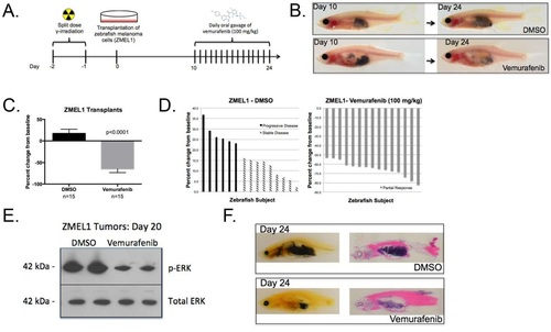
BRAFV600E inhibitor treatment of zebrafish melanoma cell line (ZMEL1) transplants. (A) Experimental workflow: MHC caspers were exposed to 30Gy split-dose of sub-lethal irradiation day -2 and -1 prior to transplantation on day 0. MHC caspers were transplanted with 500,000 zebrafish melanoma cells (ZMEL1) and allowed a 10-day period for melanoma engraftment and proliferation. A two-week regimen of daily oral gavage of DMSO-control or 100mg/kg Vemurafenib began on day 10. (B) The top panel shows a representative ZMEL1-transplanted zebrafish treated with DMSO control over a two-week treatment regimen. The bottom panel shows a representative ZMEL1-transplanted zebrafish treated with 100mg/kg Vemurafenib over a two-week treatment regimen. (C) Average percent change from baseline of tumor area based on pigmentation in a cohort with n=15 in each treatment arm. Two-tailed paired t-test was performed for statistical analysis. Data represented as mean±s.d. of three replicates. (D) Waterfall plot depicting the range of response for the experimental cohorts, DMSO and Vemurafenib. The response was quantified by percent change of tumor area from baseline. (E) ZMEL1 tumors from DMSO control (lanes 1 and 2) and Vemurafenib-treated (lanes 3 and 4) cohorts were isolated at day 20. MAPK activity was measured via phosphorylated-ERK with total ERK as a loading control. (F) Paraffin section (left) and H&E stain (right) of representative zebrafish within a DMSO (top panel) or Vemurafenib (bottom panel) cohort at day 24.
|
