
Fig. 9
- ID
- ZDB-FIG-160726-23
- Publication
- Lagman et al., 2016 - Evolution and expression of the phosphodiesterase 6 genes unveils vertebrate novelty to control photosensitivity
- Other Figures
- All Figure Page
- Back to All Figure Page
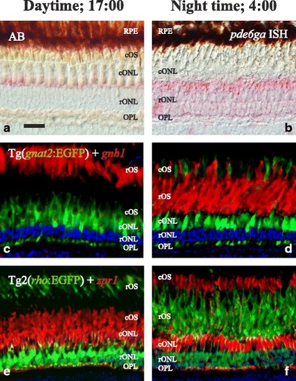
Retinomotor movements in the zebrafish retina. Photomicrographs of adult zebrafish retinae showing morphological differences between day (17:00; left column) and night (4:00; right column). In the upper row, bight field photomicrographs taken using Nomarski contrast show the clear stratification during daytime (a), different to the poorly stratified retina at night (b). In both pictures, Fast Red staining, in pink, shows the presence of pde6ga mRNAs in rods. Note the different intensity in staining, confirmed by qRT-PCR as higher expression at 4:00 AM (Fig. 7). c-f pictures are fluorescence photomicrographs of immunostainings illustrating the retinomotor movements with photoreceptor-specific markers. In c and d, sections of a transgenic line that expresses EGFP in cones: Tg(gnat2:EGFP) were incubated with a rod-specific anti-GNB1 antibody (in red). In e and f, sections of a transgenic line that expresses EGFP in rods: Tg2(rho:EGFP) were incubated with a double cone-specific anti-zpr1 antibody (in red). In both cases, note the position of the rod outer segments in the outermost part of the retina at daytime, while at night they have moved to inner positions and the cone outer segments have moved outwards. DAPI was used as a nuclei counterstain (c-f). RPE: retinal pigment epithelium. For more abbreviations, see Fig. 5 legend. Scale bar is 20 µm |