
Fig. 3
- ID
- ZDB-FIG-160714-18
- Publication
- Richardson et al., 2016 - Re-epithelialization of cutaneous wounds in adult zebrafish uses a combination of mechanisms at play during wound closure in embryonic and adult mammals
- Other Figures
- All Figure Page
- Back to All Figure Page
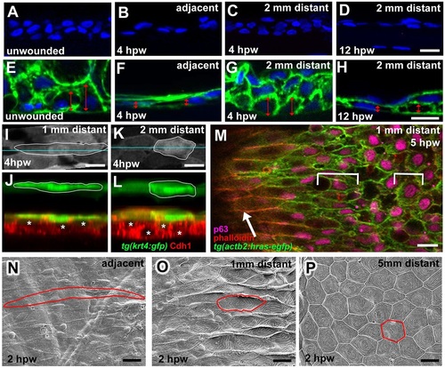
Epidermal cells outside full-thickness wounds undergo progressive radial intercalation, flattening and elongation. (A-D) DAPI labelling of sections through unwounded epidermis (A) and epidermis at indicated stages and distances from wound (B-D). (E-H) DAPI labelling of sections through Tg(actb2:hras-egfp) fish as in A-D. Cell membranes are in green. Doubled-headed red arrows indicate the heights of individual basal keratinocytes. (I-L) Surface views (I,K) and z-projections (J,L) of Tg(krt4:GFP) fish; at 1mm distance, superficial cells (outlined; green) display more pronounced flattening, and inner keratinocytes (E-cadherin; red; centres indicated by asterisks) more pronounced radial intercalations than at 2mm distance from wound. (M) Single-plane confocal micrograph of Tg(actb2:hras-egfp) fish; superficial cells are labelled with Phalloidin (red), inner epidermal cells with p63 (pink) and cell membranes with GFP (green); wound is to the left. Arrow indicates superficial cells, brackets indicate lengths of inner keratinocytes, which are less elongated than superficial cells. (N-P) SEM images of superficial skin layer (single cells outlined) at indicated distances from wound, revealing spatially graded cell elongation and loss of surface microridges. Scale bars: 10µm. |
| Genes: | |
|---|---|
| Antibodies: | |
| Fish: | |
| Condition: | |
| Anatomical Terms: | |
| Stage: | Adult |