
FIGURE
Fig. S2
Fig. S2
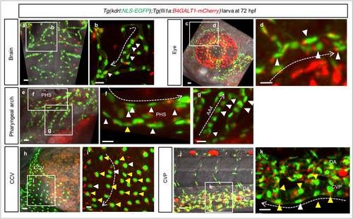
Endothelial cell polarization in different vascular beds. (a-k) 3D-rendered confocal stack images of different vascular beds: brain (a, b), eye (c, d), pharyngeal arch (e-g), CCV (h, i) and CVP (j, k) of a 72 hpf Tg(kdrl:NLS-EGFP);Tg(fli1a:B4GALT1-mCherry) larva. White arrowheads point to polarized ECs, yellow arrowheads to non-polarized ECs. Scale bars, 15 µm. White dashed arrows indicate the direction of blood flow. Anterior to the left, dorsal to the top. AA4, secondary pharyngeal arch; CCV, common cardinal vein; CVP, caudal vein plexus; DA, dorsal aorta; PHS, primary head sinus. |
Expression Data
Expression Detail
Antibody Labeling
Phenotype Data
Phenotype Detail
Acknowledgments
This image is the copyrighted work of the attributed author or publisher, and
ZFIN has permission only to display this image to its users.
Additional permissions should be obtained from the applicable author or publisher of the image.
Full text @ Nat. Commun.