
Fig. S1
- ID
- ZDB-FIG-160607-4
- Publication
- Enyedi et al., 2016 - The Cell Nucleus Serves as a Mechanotransducer of Tissue Damage-Induced Inflammation
- Other Figures
- All Figure Page
- Back to All Figure Page
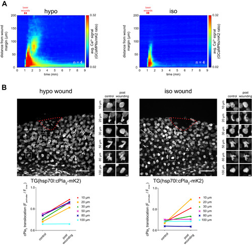
Spatiotemporal Mapping of Wound-Induced Ca2+ Signals and cPla2 Translocation in Live Zebrafish, Related to Figures 1 and 2 (A) Average spatiotemporal Ca2+ signal profile of the indicated number of transgenic Ca2+ reporter larvae Tg(hsp70l: GCaMP6s-NLS-P2A-mK2-NLS) after wounding in hypotonic (left) or isotonic (right) solution. (B) Top panels, representative confocal images showing cPla2-mK2 localization in Tg(hsp70l:cPla2-mK2) larvae after laser-wounding (wound margin marked by red dashed line) under hypotonic (left) or isotonic (right) conditions. Bottom panel, quantification of cPla2-mK2 INM translocation state in the cells selected at various distances from the wound margin. |
Reprinted from Cell, 165, Enyedi, B., Jelcic, M., Niethammer, P., The Cell Nucleus Serves as a Mechanotransducer of Tissue Damage-Induced Inflammation, 1160-1170, Copyright (2016) with permission from Elsevier. Full text @ Cell