
Fig. 7
- ID
- ZDB-FIG-160607-1
- Publication
- Li et al., 2016 - Ubr3, a Novel Modulator of Hh Signaling Affects the Degradation of Costal-2 and Kif7 through Poly-ubiquitination
- Other Figures
- All Figure Page
- Back to All Figure Page
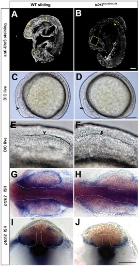
Ubr3 is required for Hh signaling and proper optic vesicle morphogenesis. (A-B) Lateral views of zebrafish embryos at 18-somites stage after removing the yolks, labeled with anti-Ubr3 antibody. Boxes outline the zebrafish retina, arrow points to central nervous system. The remaining signal in ubr3b1250/b1251 mutant corresponds to non-specific autofluorescence emanating from the yolk and some peridermal cells (arrowheads). (C-D) DIC (Differential Interference Contrast) imaging showing lateral views of 6-somites stage zebrafish embryos. (C) Wild-type optic vesicle (arrowheads) is morphologically visible and characterized by a stratified epithelial thickening. The cavitation seam divides the presumptive retina into dorsal and ventral halves. (D) ubr3 trans-heterozygous mutant optic vesicle (arrow) has morphological defects characterized by disorganized tissue that lacks epithelial morphology. The cavitation seam is disrupted or absent. (G-J) In situ hybridization for ptch2 in wild-type (G, I) and ubr3 mutant (H, J) zebrafish embryos. Note the down-regulation of ptch2 in ubr3 mutant optic vesicles (dotted area). Dorsal views of flat-mounted zebrafish embryos. (G, H) Cross sections through the optic vesicle of embryos shown in G and H, respectively. All the scale bars represent 50µm. |
| Gene: | |
|---|---|
| Antibody: | |
| Fish: | |
| Anatomical Terms: | |
| Stage: | 14-19 somites |
| Fish: | |
|---|---|
| Observed In: | |
| Stage Range: | 5-9 somites to 14-19 somites |