
Fig. 4
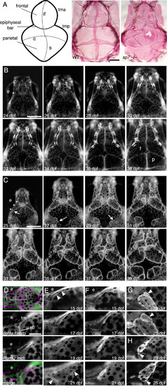
Multiple skull bones in sp7 mutants arise from independent ossification sites. (A) Schematic diagram of the skull showing the flat bones (frontal and parietal), the sutures and the cartilage bars. And alizarin red stained dissected skulls from WT and sp7 mutant fish at 6 wpf. In WT, the vault of the skull is made up of paired frontal and parietal bones that overlap by ~6 weeks to form the interfrontal, coronal, and sagittal sutures. sp7 mutants have multiple irregular bones that fail to overlap and form normal sutures. (B and C) We followed skull formation dynamically by imaging individual Runx2:egfp transgenic fish every 2 days. In a WT fish (B), the frontal and parietal bones normally form as single ossifications. The position where the initiation site grows from is indicated (asterisks). In an sp7 mutant (C), additional ossification sites arise laterally (arrows) and grow apically, giving rise to separate bones. Three initiation sites are shown with asterisks. To examine initiation of the frontal bone, we imaged transgenic fish from the earliest appearance of precursor cells. The taenia marginalis is positioned on the top of the figures (D-G). In fish doubly transgenic for a cartilage marker (col1a1:egfp) and Runx2:mch, we first observe precursor cells closely associated with the taenia marginalis anterior cartilage, just posterior to the epiphyseal bar, dashed square area (D). In WT fish (E), the cells expand in 6 days from a thin line to outline the wedge-shaped frontal bone (arrowheads). In sp7 mutants over the same period (F), the cells instead cluster along the cartilage, and stay more closely associated during early growth. G and H illustrate from two different fish the genesis of ectopic bones (arrowheads), starting from small clusters of cells adjacent to the initial ossifications. Scale bars for A-C are 1 mm, and for D–H 0.5 mm. Abbreviations: coronal (c), epiphyseal bar (eb), eye (e), frontal (f), interfrontal (if), parietal (p), sagittal (s), taenia marginalis anterior (tma), tania marginals posterior (tmp). |
| Fish: | |
|---|---|
| Observed In: | |
| Stage Range: | Days 21-29 to Days 30-44 |
Reprinted from Developmental Biology, 413(2), Kague, E., Roy, P., Asselin, G., Hu, G., Stanley, A., Albertson, C., Simonet, J., Fisher, S., Osterix/sp7 limits cranial bone initiation sites and is required for formation of sutures, 160-72, Copyright (2016) with permission from Elsevier. Full text @ Dev. Biol.