
Fig. 2
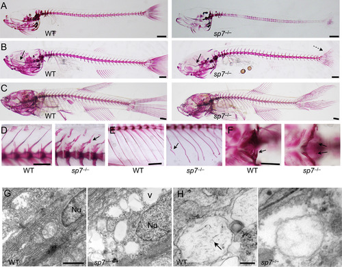
sp7 mutants show progressive skeletal malformations and deficient mineralization. (A-F) Alizarin red stain was performed on sp7 mutants and WT siblings from 2 wpf to 8 wpf. At 2 wpf (A), the skeleton of mutants shows an overall delay in mineralization. The vertebral column is unevenly mineralized and the vertebrae weakly stained. At 4 wpf (B), mineralization in the vertebral column is similar in WT and mutant. However, the skulls of mutants start to manifest a domed shape, the parasphenoid is bent (arrow), and the caudal fin lepdotrichia are wavy (dashed arrow). At 6 wpf (C), midface hypoplasia, protruding mandible and domed skull are evident in mutants. (D) Higher magnification shows the differences in size and shape of vertebrae between WT and mutant fish at 6 wpf. Arrow indicates bending of the dorsal spinous process of a single vertebra. (E) Also at 6 wpf, ribs of mutants frequently show evidence of fractures (arrow). (F) At 4 wpf, the pharyngeal teeth in the fifth ceratobranchial are fully mineralized in WT; while present in the mutants, only the tips are mineralized (arrows). (G and H) Transmission electron microscopy was performed on parietal bones at 6 wpf. (G) The nucleus (Nu) of a normal mature osteoblast is elongated and filled with dense material. In sp7 mutants the nuclei are rounder and filled with a less dense material. The extracellular matrix of mutant bone contains empty vacuoles (v), not observed in WT. (H) Needle-shaped hydroxyapatite crystals are present inside extracellular matrix vesicles in WT bone (arrow), but not observed in sp7 mutants. Scale bars for A-F are 5 mm, and for G and H are 500 µm and 100 µm, respectively. |
Reprinted from Developmental Biology, 413(2), Kague, E., Roy, P., Asselin, G., Hu, G., Stanley, A., Albertson, C., Simonet, J., Fisher, S., Osterix/sp7 limits cranial bone initiation sites and is required for formation of sutures, 160-72, Copyright (2016) with permission from Elsevier. Full text @ Dev. Biol.