
Fig. S2
- ID
- ZDB-FIG-160524-26
- Publication
- Ohnmacht et al., 2016 - Spinal motor neurons are regenerated after mechanical lesion and genetic ablation in larval zebrafish
- Other Figures
- All Figure Page
- Back to All Figure Page
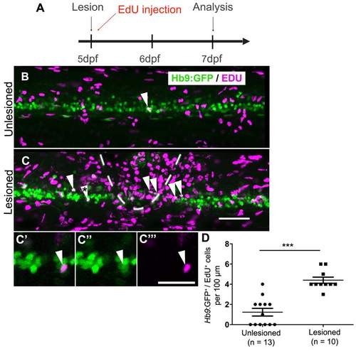
In older larvae, a lesion also leads to motor neuron regeneration within 48 hours. (Lateral views are shown; rostral is left, dorsal is up. The lesion site is indicated by dashed line) A: The timeline of the experiment is shown. B,C: The density of EdU labeled Hb9:GFP+ motor neurons (arrowheads) is strongly increased in the vicinity of a lesion site (dashed line in B). C′-C′′: The double-labeled cell marked by an asterisk in C is shown at higher magnification in a single optical section. D: Quantification shows a significant increase in motor neuron generation between 5 and 7 dpf (t-test, ***P < 0.0001). Scale bar C = 50 µm for B,C; in C′′′ = 20 µm for C′-C′′′. |