
Fig. 6
- ID
- ZDB-FIG-160428-14
- Publication
- Sallin et al., 2016 - Acute stress is detrimental to heart regeneration in zebrafish
- Other Figures
- All Figure Page
- Back to All Figure Page
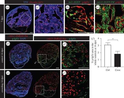
Daily stress decreases phospho-Igf1r activation in the regenerating heart. (a) Ventricular sections at 7 dpci reveal the presence of phospho-Igf1r (p-Igfr1, red) in the endocardium that covers cardiac muscle labelled by tropomyosin expression (TMP, blue). (b) p-Igf1r partially colocalizes with the pattern of endothelial cell layers demarcated by GFP expression in tie2::EGFP transgenic fish. (c) p-Igf1r does not overlap with the plasma membrane of CMs in cmlc2::EGFP-PM transgenic fish. (d,e) Sections of cmlc2::DsRed2-Nuc (red) hearts at 14 dpci display reduced p-Igf1r (green) expression in stressed animals. (f) Quantification of phospho-Igf1r area per ventricle at 14 dpci in control and stressed animals (N ≥ 7). *p < 0.05. Dashed lines encircle the cryoinjured areas. Scale bars, 100 µm. |
| Gene: | |
|---|---|
| Antibody: | |
| Fish: | |
| Anatomical Term: | |
| Stage: | Adult |