
Fig. 4 S2
- ID
- ZDB-FIG-160321-21
- Publication
- Eom et al., 2015 - Long-distance communication by specialized cellular projections during pigment pattern development and evolution
- Other Figures
- All Figure Page
- Back to All Figure Page
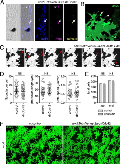
Expression of dnCdc42 transgene for inhibition of airinemes.(A) Trunk cross-section during pigment pattern formation (~7.5 SSL) for individual expressing aox5:Tet:nVenus-2a-dnCdc42 mosaically and stained for Venus immunoreactivity. Nuclear-localizing Venus was co-expressed with xanthophore lineage marker Pax7 (Minchin and Hughes, 2008) (arrow; arrowhead indicates a Pax7+ cell not carrying the transgene). (B) High-resolution micrograph of aox5+ xanthoblast in wild-type zebrafish (7.5 SSL) illustrating airineme with vesicle (arrow) as well as short filopodia that lack vesicles (e.g., arrowhead). (C) Frames from time-lapse imaging (5 min intervals) showing aox5+ cells after several days of dd treatment to express dnCdc42 at low levels. Short filopodia (e.g., arrowheads) and other protrusions (e.g., *) were extended despite inhibition of airineme production. (D) Analyses of protrusive activities in time-lapse videos revealed no significant differences in numbers of short filopodia extended (left: t58=1.07, p=0.3), lengths of lamellipodial protrusions (center: t54=1.20, p=0.2), or speeds of lamellipodial extension (right: t54=0.62, p=0.4). (E) Following extended dd-induction of dnCdc42 (6.5–12.5 SSL), there were not significant differences in the total numbers of xanthophores (t8=0.83, p=0.4) or melanophores (t8=0.27, p=0.8) relative to dd-treated non-transgenic controls. (F) Distributions of aox5+ cells were similar between dd-treated dnCdc42 and control larvae. Scale bars: 20 µm (A); 5 µm (C); 20 µm (C); 50 µm (F). |