
Fig. 7
- ID
- ZDB-FIG-160317-8
- Publication
- Lisse et al., 2016 - Comparative transcriptomic profiling of hydrogen peroxide signaling networks in zebrafish and human keratinocytes: Implications toward conservation, migration and wound healing
- Other Figures
- All Figure Page
- Back to All Figure Page
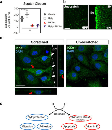
(a) IKK is necessary for H2O2-induced HEK01 keratinocyte migration after wounding. Scratch assays were performed with H2O2 (0.1 µM) and Wedelolactone (50 µM) using HEK01 cells. Two-way ANOVA at an alpha = 0.05 (95% confidence interval) and Bonferroni’s multiple comparison post-tests were utilized. Significance is denoted with asterisks: *p < 0.05, **p < 0.01 (n ≥ 3-5 cell culture experiments). (b) Rapid H2O2 production using 1 µM HPF (hydrogen peroxide fluorogenic probe) at the scratch (sc) margin of HEK01 keratinocytes within 30 minutes. Bar = 100 µm (c) Rapid subcellular accumulation of IKKα within injured HEK01 cells at the scratch (sc) margin (white arrows) after 30 minutes compared to unscratched cells. Orthogonal views (red arrows) of an injured cell show peri-nuclear and cytoplasmic distribution and accumulation of IKKα. Bar = 20 µm. (d) Schema of overall findings. |