
Fig. 2
- ID
- ZDB-FIG-160317-6
- Publication
- Lisse et al., 2016 - Comparative transcriptomic profiling of hydrogen peroxide signaling networks in zebrafish and human keratinocytes: Implications toward conservation, migration and wound healing
- Other Figures
- All Figure Page
- Back to All Figure Page
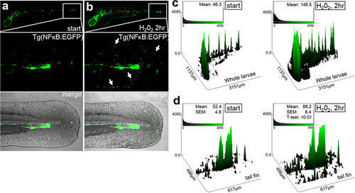
Transgenic NF-κB:EGFP zebrafish reveal peripheral NF-κB activation after H2O2 treatment Spatial patterns of increased NF-κB activation after H2O2 treatment of zebrafish larvae. (a) 3-dpf Tg(NF-κB:EGFP) reporter zebrafish larvae at the start of the experiment. (b) Tg(NF-κB:EGFP) zebrafish were imaged after 2hr post 0.01% H2O2 treatment. Both whole larvae (upper) and tail fins (below) were imaged, whereby GFP+ cells were partially localized to the periphery (arrows). (c) Quantitative analysis using relative mean fluorescence of the z-stack projected images using ImageJ (n = 2, 10 fish). Observation of increased number of GFP-labeled cells and overall fluorescence intensity in the whole larvae. (d) Higher-resolution analyses of the tail fin revealed a peripheral tissue spatial pattern of increase NF-κB activated cells after H2O2 treatment. n = 3 independent experiments per condition. |
| Gene: | |
|---|---|
| Fish: | |
| Condition: | |
| Anatomical Term: | |
| Stage: | Protruding-mouth |
| Fish: | |
|---|---|
| Condition: | |
| Observed In: | |
| Stage: | Protruding-mouth |