
Fig. 2
- ID
- ZDB-FIG-160222-7
- Publication
- Macdonald et al., 2016 - Assessment of biocompatibility of 3D printed photopolymers using zebrafish embryo toxicity assays
- Other Figures
- All Figure Page
- Back to All Figure Page
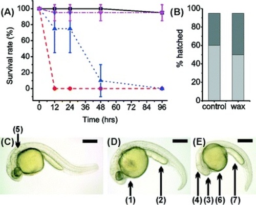
(A) Survival rate of zebrafish embryos in 3D printed material VisiJet Crystal without S300 support material (red circles) and with (dark blue triangle). Zebrafish cultured within a Petri dish containing S300 (magenta inverted triangle) and without as a control (black square). Error bars span one standard deviation from the mean (B) hatching of zebrafish embryos at 48 h (light grey) and 96 h (dark grey), in a Petri dish containing S300 (‘wax’) and without (‘control’). (5 embryos per well, n = 5). (C–E) Morphology analysis at 27 hours incubation of zebrafish with 3D printed materials. Stereomicroscopy images of dechorionated zebrafish embryos immobilised in agar: (C) control zebrafish embryo cultured in a 24-well plate development is normal. (D) Image of zebrafish cultured with Watershed material. Development has been stunted, appears 2–3 hours behind control, darkening of yolk sac (1) indicating toxicity as well as roughing and widening yolk extension (2). (E) Image of zebrafish cultured on VisiJet Crystal. Development has been stunted by ca. 5 hours. Eyes (3) and brain (4) have not developed, pigmentation (shown as (5) in C) is not present, unusual yolk sac (6) and yolk extension (7) shape, all show retardation. Scale bar is 500 µm. |