
Fig. 1
- ID
- ZDB-FIG-160212-30
- Publication
- Nicolson, 2015 - Ribbon Synapses in Zebrafish Hair Cells
- Other Figures
- All Figure Page
- Back to All Figure Page
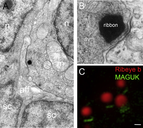
Ribbon synapses in zebrafish hair cells. A, Transmission electron microscopy (TEM) micrograph of the basal end of an inner ear hair cell located within the anterior macula. A prominent ribbon with vesicles is seen juxtaposed to an afferent fiber that appears to contact a large surface area (a second faint ribbon outside of the sectioning plane is seen to right). B, High magnification view of a ribbon along with the postsynaptic density. C, Super resolution structured illumination microscopy (SR-SIM) image of several ribbon synapses labeled with Ribeye b (red) and pan-MAGUK (green) antibodies (Image: Lavinia Sheets). Abbreviations: aff, afferent; m, mitochondria; n, nucleus; PSD, postsynaptic density; sc, supporting cell; SV, synaptic vesicle. Scale bar, 200 nm in A; 75 nm in B; C, 250 nm. |
Reprinted from Hearing Research, 330(Pt B), Nicolson, T., Ribbon Synapses in Zebrafish Hair Cells, 170-7, Copyright (2015) with permission from Elsevier. Full text @ Hear. Res.