
Fig. 2
- ID
- ZDB-FIG-160208-58
- Publication
- Enright et al., 2015 - Cyp27c1 Red-Shifts the Spectral Sensitivity of Photoreceptors by Converting Vitamin A1 into A2
- Other Figures
- All Figure Page
- Back to All Figure Page
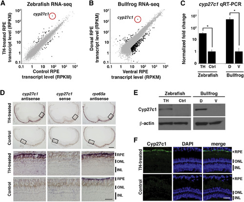
Cyp27c1 Expression Is Correlated with the Presence of Vitamin A2 in Zebrafish and Bullfrog RPE
(A) Zebrafish were treated with TH or a vehicle control for 3 weeks, after which RPE was isolated and used to construct a cDNA library for transcriptome profiling by RNA-seq. Expression levels (in RPKM [reads per kilobase of transcript per million mapped reads]) of individual transcripts from TH-treated RPE (y axis) and vehicle-treated RPE (x axis) are shown as dots, with significantly differentially expressed genes in black (quantile-adjusted conditional maximum likelihood [qCML] test; FDR < 0.05; n = 3). (B) Dorsal and ventral thirds of bullfrog RPE were isolated and used to construct a cDNA library for transcriptome profiling by RNA-seq. Expression levels of individual transcripts from dorsal RPE (y axis) and ventral RPE (x axis) are shown as dots, with significantly differentially expressed genes in black (qCML test; FDR < 0.05; n = 3). (C) Enrichment of the cyp27c1 transcript in cDNA samples used for RNA-seq was confirmed by quantitative real-time PCR (qRT-PCR). Expression was normalized to ribosomal protein rpl13a for zebrafish and rpl7a for bullfrog (two-sided Student’s t test; n = 2–3; p < 0.005; error bars = SEM). (D) In situ hybridization of albino zebrafish treated with TH or vehicle control for 3 weeks. Top panels show cross-sections of the whole eye. Bottom panels show high-magnification images of the boxed regions from the top panels. The antisense probe for cyp27c1 localized exclusively to the RPE in TH-treated fish. No signal was observed with the cyp27c1 sense probe, but strong signal was observed in the RPE of TH-treated and control fish with an antisense probe against rpe65a, a gene that is expressed at high levels in RPE. The scale bars represent 200µm low power and 50 µm high power. (E) Western blot with a rabbit polyclonal anti-Cyp27c1 antibody confirmed enrichment of Cyp27c1 protein in TH-treated zebrafish and dorsal bullfrog RPE. β-actin was used as a loading control. (F) Immunohistochemistry of albino TH- and vehicle-treated zebrafish indicates induction of Cyp27c1 expression in the RPE of TH-treated fish (green), with DAPI used to counter-stain nuclei (blue). The scale bar represents 50 µm. See also Figure S1 and Tables S1 and S2. |