
Fig. 1
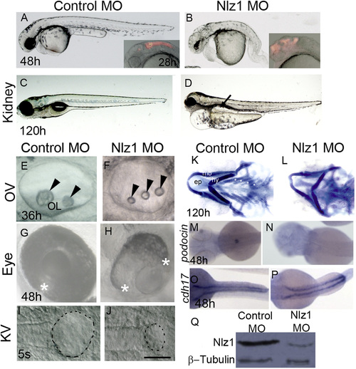
Nlz1 morpholino-injected embryos show morphological defects similar to a human ciliopathy. (A)–(J) DIC images of live embryos at different developmental stages (A), (B), (G), and (H) 48 h; (C), and (D) 120 h; (E), (F) 36 h; (I), and (J) 5S. Embryos were injected with Control MO (A), (C), (E), (G), and (I) or with Nlz1 MO (B), (D), (F), (H), (J) at the 1–2 cell stage. Nlz1 MO injected embryos show generalized edema; an abnormal brain; a short, bent tail (B); kidney cyst(s) (D, arrow, n=45/55); three small otoliths in the OV (F, arrow head, n=52/60); and a rudimentary KV (J, dotted line, n=15/40). In control MO injected embryos, gross morphology (A), kidneys (C), otoliths (E), and KV (I) were normal. Control MO injected embryos (A, inset), and nlz1 morphants (B, inset) were injected with rhodamine dextran in the hindbrain ventricles at 28 h.The two edges of the optic fissure were fused completely in control MO injected embryos (G, asterisk, n=50/50) whereas coloboma was observed in nlz1 morphants (H, n=72/80). (K)–(L) Alcian blue staining of pharyngeal cartilages in control MO (K) and Nlz1 MO (L) injected zebrafish larvae at 120 h. (M)–(P) Dorsal view of a whole mount in situ hybridization with probes for podocin (M), and (N), and cdh17 (O), and (P) in control MO (M), and (O) and Nlz1 MO (N), and (P) injected embryos at 48 h. (Q) Western blot shows severe reduction of Nlz1 protein expression in morphants. (B), (F), (H), and (J) injected with 3.5 ng Nlz1 MO, (D), (L), (N), and (P) injected with 2 ng Nlz1 MO. (A)–(H) lateral views, anterior to the left; (I)–(J) dorsal view, posterior to the top. Scale bar in (A) and (B), and insets, 320 µm; (C) and (D), 200 µm; (E) and (F), 35 µm; (G) and (H), 100 µm; (I) and (J), 50 µm. OV, otic vesicle; KV, Kupffer’s vesicle; OL, otolith; s, somite; ep, ethmoid plate; mb, mandibular arch; hy, hyoid arch; asterisks indicate branchial arches. |
| Genes: | |
|---|---|
| Fish: | |
| Knockdown Reagent: | |
| Anatomical Terms: | |
| Stage: | Long-pec |
| Fish: | |
|---|---|
| Knockdown Reagent: | |
| Observed In: | |
| Stage Range: | 5-9 somites to Day 5 |
Reprinted from Developmental Biology, 406(2), Dutta, S., Sriskanda, S., Boobalan, E., Alur, R.P., Elkahloun, A., Brooks, B.P., nlz1 Is required for cilia formation in zebrafish embryogenesis, 203-11, Copyright (2015) with permission from Elsevier. Full text @ Dev. Biol.