
Fig. 4
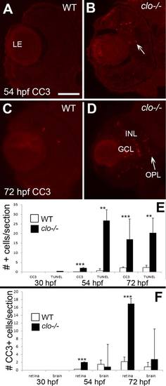
Increased retinal cell death during late retinal neurogenesis in clo mutant embryos. A–D: Immunofluorescence images of wild-type (A,C) and clo-/- (B,D) retinas stained with anti-cleaved caspase 3 (CC3); samples obtained at 54 hpf (A,B) and 72 hpf (C,D). Arrow in B indicates examples of CC3+ cells. E: Numbers of CC3+ and TUNEL+ cells are significantly (***P < 0.001; **P < 0.01) increased in clo-/- retinas at 54 hpf and 72 hpf. F: Numbers of CC3+ (dying) cells are significantly (***P < 0.001) increased in clo-/- retinas at 54 hpf and 72 hpf, but not in clo-/- brains. Scale bar = 50 µm in A (applies to all images); LE, lens; GCL, ganglion cell layer; INL, inner nuclear layer; OPL, outer plexiform layer. |