
Fig. 3
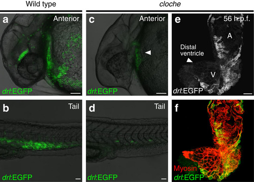
Lack of endocardial signals does not affect integration of FHF with SHF. (a,b) Anterior (a) and tail region (b) expression of drl:EGFP in wild-type zebrafish at 24–28 h.p.f., scale bar, 50 µm; note the circulating red blood cells. (c,d) Anterior (c) and tail region (d) drl:EGFP expression at 24–28 h.p.f. in the recessive zebrafish mutant cloche, which lacks all endothelial and haematopoietic lineages; scale bar, 50 µm; note the formation of linear heart tube in cloche (solid arrowhead in c and residual angioblasts in the tail (d). (e,f) Top-down 2-µm confocal projection of 56 h.p.f. cloche mutant heart, drl:EGFP expression counter stained with anti-GFP (e, green in f) and cardiomyocytes stained with myosin/MF20 (red in f); scale bar, 50 µm; note how despite missing endocardium the heart features both demarcated FHF and SHF myocardium, solid arrowhead in e indicates the distal ventricle region. Atrium (A) and ventricle (V) labelled in e. |