
Fig. S2
- ID
- ZDB-FIG-151008-4
- Publication
- Aspatwar et al., 2015 - Inactivation of ca10a and ca10b Genes Leads to Abnormal Embryonic Development and Alters Movement Pattern in Zebrafish
- Other Figures
- All Figure Page
- Back to All Figure Page
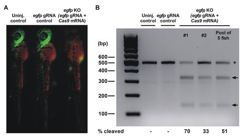
Silencing of egfp expression in tg(fli1a:egfp) zebrafish embryos using CRISPR/Cas9 mediated mutagenesis. A) Fluorescence microscopy was used to analyze the silencing of egfp in tg(fli1a:egfp) zebrafish embryos at 2 dpf. The un-injected control (left) and egfp gRNA injected control (middle) express egfp (green) in the vascular endothelium. The CRISPR-Cas9 mutated embryo (right) shows less fluorescence due to the disruption of the egfp gene. The red channel was used to detect auto-fluorescence. B) T7 endonuclease I (T7EI) assay was used to evaluate the egfp mutation efficiency in 2-day-old embryos. T7EI treated PCR products of un-injected and egfp gRNA injected control fish are shown in comparison to PCR products of two individual embryos and a pooled sample of 5 egfp silenced embryos. The full length wild type (WT) egfp product (470bp) is marked with an asterix. Arrows indicate the T7E1 cleaved PCR products in the egfp mutated embryos. |