
Fig. 4
- ID
- ZDB-FIG-151001-1
- Publication
- Giousoh et al., 2015 - Bone morphogenetic protein/retinoic acid inducible neural-specific protein (brinp) expression during Danio rerio development
- Other Figures
- All Figure Page
- Back to All Figure Page
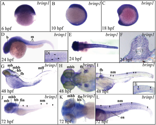
The spatial expression pattern of brinp1 during zebrafish development. brinp1 is broadly expressed between 6 hpf and 16 hpf (A–C) and is detected in the brain (b) and trunk muscle (m) at 24 hpf (D), close up view is shown of the myosepta (Di). Cross section at 24 hpf shows staining in the muscle (F). At 48 hpf, brinp1 is specifically detected in the midbrain (mb), mid-hindbrain (mhb), hindbrain (hb), fin buds (fb), median fin fold (mff) and lateral line neuromasts (nm) (E–G). A similar expression pattern is observed at 72 hpf. Views are lateral with anterior to the left (D, G, I, J, L) and dorsal views are shown in E, H and K. |
| Gene: | |
|---|---|
| Fish: | |
| Anatomical Terms: | |
| Stage Range: | Shield to Protruding-mouth |
Reprinted from Gene expression patterns : GEP, 18(1-2), Giousoh, A., Vaz, R., Bryson-Richardson, R.J., Whisstock, J.C., Verkade, H., Bird, P.I., Bone morphogenetic protein/retinoic acid inducible neural-specific protein (brinp) expression during Danio rerio development, 37-43, Copyright (2015) with permission from Elsevier. Full text @ Gene Expr. Patterns