
FIGURE
Fig. 1
- ID
- ZDB-FIG-150929-36
- Publication
- Rajaram et al., 2014 - Technical brief: Constant intense light exposure to lesion and initiate regeneration in normally pigmented zebrafish
- Other Figures
- All Figure Page
- Back to All Figure Page
Fig. 1
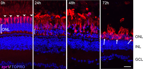
Effects of constant intense light exposure on cone cells of pigmented AB zebrafish. Retinas were collected from zebrafish before (0 h) and after the indicated length of light exposure. Retinas were processed for immunohistochemistry and stained with zpr1 antibody (red). Nuclei were counter-stained with TOPRO (blue). Dorsal retinas are shown. The arrowheads at 0 h indicate the ordered arrangement of the double cones; the bracket indicates ONL. White arrows show condensed zpr+ cells at 72 h of intense light. ONL, outer nuclear layer; INL, inner nuclear layer; GCL, ganglion cell layer. Scale bar is 50 µm. |
Expression Data
| Antibody: | |
|---|---|
| Fish: | |
| Condition: | |
| Anatomical Terms: | |
| Stage: | Adult |
Expression Detail
Antibody Labeling
Phenotype Data
| Fish: | |
|---|---|
| Condition: | |
| Observed In: | |
| Stage: | Adult |
Phenotype Detail
Acknowledgments
This image is the copyrighted work of the attributed author or publisher, and
ZFIN has permission only to display this image to its users.
Additional permissions should be obtained from the applicable author or publisher of the image.