
Fig. 4
- ID
- ZDB-FIG-150826-20
- Publication
- Belon et al., 2015 - A Macrophage Subversion Factor Is Shared by Intracellular and Extracellular Pathogens
- Other Figures
- All Figure Page
- Back to All Figure Page
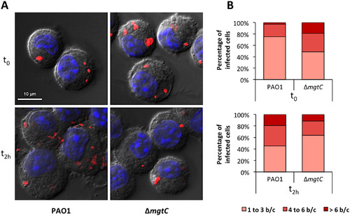
Visualisation and quantification of intracellular bacteria in fixed macrophages. Macrophages infected with PAO1 or ΔmgtC mutant expressing mCherry were fixed after phagocytosis and 20 min (t0) or 2 hours (t2h) treatment with amikacin. (A) Visualization of intracellular bacteria. The images shown were obtained by making a maximum projection of 12 central planes from a Z-stack. The merged image shows Differential Interference Contrast (DIC), nucleus staining (blue) and bacteria expressing mCherry (red). (B) Count of the number of bacteria in infected macrophages from images obtained with the maximum projection. The numbers of bacteria per cell (b/c) were classified in three groups and percentage of each class is shown. Count is done from at least 20 cells and results are expressed as means from three independent experiments. |