
Fig. 4
- ID
- ZDB-FIG-150626-2
- Publication
- Xu et al., 2015 - Characterization of Tetratricopeptide Repeat-Containing Proteins Critical for Cilia Formation and Function
- Other Figures
- All Figure Page
- Back to All Figure Page
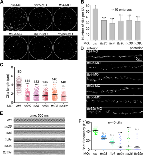
Examinations on cilia in the Kupffer’s vesicle and pronephric duct. (A-C) Both cilia number and cilia length in the Kupffer’s vesicle (KV) at the 7-somite stage were reduced in the indicated ttc morphants. Cilia were labeled using antibody against acetylated tubulin. The quantification results were based on three independent experiments. Student’s t-test, ***P < 0.001. Error bars represent s.d. (D) Typical morphology of cilia in pronephric ducts at 24 hpf. (E) and (F) Cilia motility in the pronephric duct at 60 hpf. The kymographs showed trajectories of the cilia marked with red line in S1, S2, S3, S4, S5 and S6 Videos. Cilia beat frequencies (CBFs) were measured at 60 hpf (40 cilia from 4 morphants for each gene). Student’s t-test, ***P < 0.001. Error bars represent s.e.m. |
| Fish: | |
|---|---|
| Knockdown Reagents: | |
| Observed In: | |
| Stage Range: | 5-9 somites to Pec-fin |