
Fig. 4
- ID
- ZDB-FIG-150512-4
- Publication
- Burgoyne et al., 2015 - Regulation of melanosome number, shape and movement in the zebrafish retinal pigment epithelium by OA1 and PMEL
- Other Figures
- All Figure Page
- Back to All Figure Page
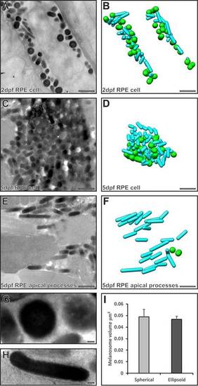
Serial section electron microscopy analysis reveals two populations of melanosomes in the RPE cell body, but only cylindrical ones enter the apical processes. (A,C,E) Single electron micrographs from a stack of serial section images used to generate models (B,D,F) of spherical (green) and cylindrical (cyan) melanosomes. (A,B) Both cylindrical and spherical melanosomes are found in the RPE cell body at 2dpf. (C,D) Cylindrical and spherical melanosomes are densely packed in the RPE cell body at 5dpf. (E,F) Only cylindrical melanosomes are in the apical processes at 5dpf. (G,H) Higher-magnification images of a spherical melanosome (G) and a cylindrical melanosome (H). Scale bars: 1µm (A–F), 100 nm (G–H). (I) Spherical and cylindrical melanosomes have similar volumes. Results show the mean±s.e.m. |