
Fig. 4
- ID
- ZDB-FIG-150507-14
- Publication
- Gramage et al., 2015 - Midkine-a Protein Localization in the Developing and Adult Retina of the Zebrafish and Its Function During Photoreceptor Regeneration
- Other Figures
- All Figure Page
- Back to All Figure Page
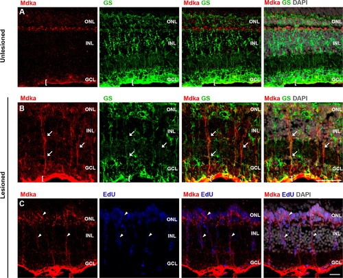
Mdka protein localization following photoreceptor ablation. In unlesioned retinas, Mdka immunostaining is localized to the horizontal cells and endfeet of glutamine synthetase (GS)-positive Müller glia (row A). At 4 dpl, Mdka antibodies label the radial processes of Müller glia (row B, arrows). Note the increased Mdka immunostaining in the endfeet of the Müller glia in lesioned retinas (cf. rows A and B). Also at 4 dpl, Mdka immunostaining is localized to each of the EdU-positive nuclei in both the INL and ONL (row C, arrowheads). ONL: outer nuclear layer; INL: inner nuclear layer; dpl: days post lesion. Scale bars = 25 µm. |
| Gene: | |
|---|---|
| Antibodies: | |
| Fish: | |
| Condition: | |
| Anatomical Terms: | |
| Stage: | Adult |
| Fish: | |
|---|---|
| Condition: | |
| Observed In: | |
| Stage: | Adult |