
Fig. 2
- ID
- ZDB-FIG-150505-11
- Publication
- Ito et al., 2014 - Differential reparative phenotypes between zebrafish and medaka after cardiac injury
- Other Figures
- All Figure Page
- Back to All Figure Page
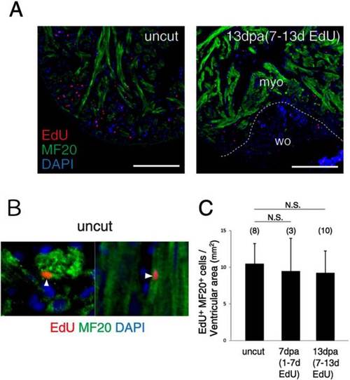
Cardiomyocyte proliferation after ventricular resection. A: Fluorescent images of EdU/ MF20 double-stained hearts. EdU-incorporating uncut and resected (13 dpa) hearts are shown (EdU incorporated from 7 to 13 dpa). B: Representative EdU/ MF20 double-positive cardiomyocyte (arrowhead) in the uncut heart. C: Quantification of EdU/MF20 double-positive cells in the ventricle. The count was normalized by the ventricular area. Numbers in parentheses indicate the sample number. Student′s t-test, N.S., not significant. The dashed line indicates the approximate injury border. myo, myocardium; wo, wound area. Scale bars = 100 µm. |