
Fig. 4
- ID
- ZDB-FIG-150428-40
- Publication
- Wang et al., 2014 - Inhibitors of neutrophil recruitment identified using transgenic zebrafish to screen a natural product library
- Other Figures
- All Figure Page
- Back to All Figure Page
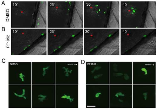
Time-lapse imaging of neutrophil migration in vivo. The ventral fin of 6-dpf larvae was injured by a sharp syringe needle, and chemotaxis of neutrophils were imaged by spinning disk microscope. (A) In a control larva treated with DMSO, neutrophil migration was not directed until 10 minutes after fin injury. Thereafter, they migrated actively towards the wound site (red asterisk; 25 and 30 minutes); by 40 minutes after injury, around eight neutrophils were recruited. (B) In contrast to the control, neutrophils treated with PF1052 remained stationary during 40 minutes of imaging; they appeared round and barely formed any pseudopods. (C) Higher magnification shows that DMSO-treated neutrophils actively formed multiple pseudopods and this was impaired in PF1052 treated-neutrophils (D). |