
FIGURE
Fig. 3
- ID
- ZDB-FIG-150427-6
- Publication
- Progatzky et al., 2013 - From seeing to believing: labelling strategies for in vivo cell-tracking experiments
- Other Figures
- All Figure Page
- Back to All Figure Page
Fig. 3
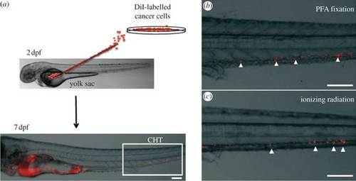
Live/dead control experimental design. (a) A549 cells labelled with CM-DiI were injected into the yolk sac of wild-type zebrafish embryos at 48 h post-fertilization and in vivo imaging was performed 5 days later. (b,c) Higher magnification images of the CHT area, where CM-DiI signal was detected despite cells being treated with toxic doses of PFA or ionizing radiation prior to injection. This level and type of CM-DiI signal therefore cannot be used to quantify micrometastasis formation. Scale bars, 100 µm. |
Expression Data
Expression Detail
Antibody Labeling
Phenotype Data
Phenotype Detail
Acknowledgments
This image is the copyrighted work of the attributed author or publisher, and
ZFIN has permission only to display this image to its users.
Additional permissions should be obtained from the applicable author or publisher of the image.
Full text @ Interface Focus