
Fig. 5
- ID
- ZDB-FIG-150408-21
- Publication
- Strzyz et al., 2015 - Interkinetic Nuclear Migration Is Centrosome Independent and Ensures Apical Cell Division to Maintain Tissue Integrity
- Other Figures
- All Figure Page
- Back to All Figure Page
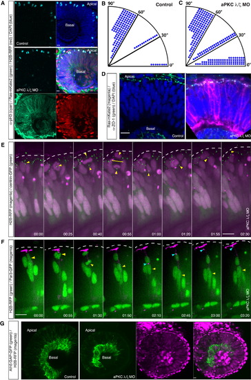
Nonperpendicular Apical Divisions Do Not Majorly Perturb Retinal Tissue Architecture and Early Neuronal Layering (A) Images of aPKC γ/ζ MO-injected embryo (middle and lower) and a control embryo (upper). Ras-mKate2 RNA (green) and H2B-RFP RNA (red) were injected to visualize morphant cells together with MOs. Embryos were fixed at 34 hpf and stained with pH3 antibody to visualize mitotic cells (cyan). In control and morphant embryos, mitoses occur apically. (B and C) Division angles of the control (B) and aPKC γ/ζ morphant cells (C). Dots indicate individual cells. In morphant cells, the clear bias for perpendicular divisions (angles 60°–90°) is lost. (B) n = 145 cells, 7 embryos; (C) n = 241 cells, 8 embryos. (D) Confocal images of a control (left) and an aPKC γ/ζ morphant embryo (right). Ras-GFP RNA (magenta) was injected together with MOs to visualize morphant cells. Embryos were fixed at 34 hpf and stained with ZO-1 antibody (green). In both cases, ZO-1 signal appears as a continuous apical belt. (E) Time-lapse of an aPKC γ/ζ morphant embryo coinjected with H2B-RFP RNA (magenta) and centrin-GFP RNA (green). Morphant cell divides horizontally (00:55, yellow bar). The more basal centrosome (yellow arrow) descends to the apical surface. Time is in hr:min. The frames are from Movie S6. See also Figure S2. (F) Time-lapse of an aPKC γ/ζ morphant embryo coinjected with H2B-RFP RNA (green) and Par3-GFP RNA (magenta). Morphant cell divides horizontally (01:30). The more basal daughter (yellow arrow) re-establishes its apical Par3 domain (cyan arrow). Time is in hr:min. The frames are from Movie S6. (G) Early neuronal layer, RGC (marked by Tg(Ath5:GAP-GFP), green) layer in a control embryo (left-most panel) and an aPKC γ/ζ morphant embryo (remaining panels). Morphant cells were marked by coinjection of H2B-RFP RNA (magenta). Both in the morphant and the control embryo an intact RGC layer (green) is formed next to the lens. Morphant cells (magenta) contribute to this layer (rightmost panel). Scale bars represent 10 µm. The dotted line represents the apical surface. |
Reprinted from Developmental Cell, 32(2), Strzyz, P.J., Lee, H.O., Sidhaye, J., Weber, I.P., Leung, L.C., Norden, C., Interkinetic Nuclear Migration Is Centrosome Independent and Ensures Apical Cell Division to Maintain Tissue Integrity, 203-19, Copyright (2015) with permission from Elsevier. Full text @ Dev. Cell