
Fig. 2
- ID
- ZDB-FIG-150408-18
- Publication
- Strzyz et al., 2015 - Interkinetic Nuclear Migration Is Centrosome Independent and Ensures Apical Cell Division to Maintain Tissue Integrity
- Other Figures
- All Figure Page
- Back to All Figure Page
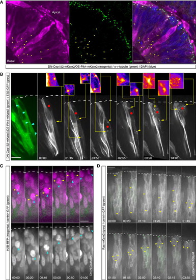
Apical IKNM Persists in Cells in which Centrosomes and Nucleus Meet Nonapically Even following Nonapical Mitotic Entry (A) Confocal scan of a HS-DN-Cep152-mKate2/HS-OS-Plk4-mKate2 expressing embryo. Positive cells feature cytosolic mKate2 signal as well as foci of signal along the apicobasal axis (magenta). Foci are positive for γ-tubulin staining (green). Nonapical foci positive for mKate2 and immunopositive for γ-tubulin are marked with yellow arrows. HS was performed 8 hr prior to fixing. See also (B) Time-lapse of a cell expressing HS-DN-Cep152-mKate2, HS-OS-Plk4-mKate2 (green), and the dynamic MTs marker HS-EB3-GFP (gray). (Left) Cytosolic mKate2 signal and nonapical foci (cyan outlined arrows) in the cell of interest are shown. The remaining panels show distribution of dynamic MTs. Clear foci of nonapical MT nucleation can be observed (yellow arrows). In the insets, magnified regions of nonapical MT nucleation sites are shown in fire lookup table (in panels 4 and 7, 1.5× magnification, remaining panels 3× magnification). The red dot marks the position of the nucleus. HS was performed 12.5 hr prior to time-lapse. Time is in hr:min. The frames are from Movie S2. (C) Time-lapse of a cell expressing DN-N-Cadherin. HS-H2B-RFP labels nuclei/chromatin (magenta in the upper and gray in the lower) and centrin-GFP-RNA labels centrosomes (green, upper panel only). The centrosome (yellow arrow) associates with the nucleus (cyan, arrow) in a nonapical position (upper). The cell enters mitosis nonapically as visualized by chromosome condensation (lower, 00:40, cyan arrow). Apical IKNM and apical division occur. HS was preformed 17.5 hr prior to time-lapse. Time is in hr:min. The frames are from Movie S2. See also Figure S2. (D) Time-lapse of a cell in an embryo treated with 100 µM colcemide. The cell expresses Ras-mKate2 (gray) and centrin-GFP (green). In the cell of interest the split centrosome (yellow arrows) travels basally and associates with the nucleus (yellow dot) nonapically. After nonapical cell rounding (01:50), the cell performs apical IKNM. Time-lapse was started 5 hr after drug addition. Time is in hr:min. The frames are from Movie S3. Scale bars represent 10 µm. The dotted line represents the apical surface. |
Reprinted from Developmental Cell, 32(2), Strzyz, P.J., Lee, H.O., Sidhaye, J., Weber, I.P., Leung, L.C., Norden, C., Interkinetic Nuclear Migration Is Centrosome Independent and Ensures Apical Cell Division to Maintain Tissue Integrity, 203-19, Copyright (2015) with permission from Elsevier. Full text @ Dev. Cell