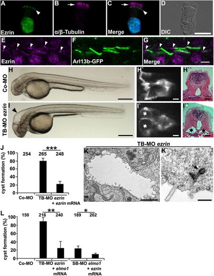
Ezrin shows expression in cilia and is required for ciliogenesis in zebrafish. (A-D) Human respiratory epithelial cells from healthy controls were double labelled with antibodies directed against Ezrin (green) and the ciliary axoneme marker α / β -Tubulin (magenta) (arrow). Ezrin localisation is restricted to the putative basal bodies (arrowhead) and the nucleus. The nucleus is stained with Hoechst 33342 (blue). (E-G) Ezrin is expressed at the basal bodies (arrowheads) and the ciliary axonemes in 48 hpf Tg(actb2:Mmu.Arl13b-GFP) zebrafish embryos. (H-I′′) Expression silencing of ezrin (I-I′′) using TB-MO ezrin (2 ng) results in hydrocephalus (arrowhead in I) and pronephric cyst formation (stars in I′ and I′′) as compared with zebrafish embryos injected with Co-MO (2 ng) (H-H′′), shown in a bright-field lateral view with anterior to the left (H,I), a dorsal view with anterior to the left of a Tg(wt1b:EGFP) embryo (H′,I′), and by a histological transverse section (H′′,I′′) of 48 hpf embryos. (J) Quantification of pronephric cyst formation in 48 hpf zebrafish embryos after injection with TB-MO ezrin (2 ng) or TB-MO ezrin (2 ng) + ezrin mRNA (20 pg), as compared with Co-MO (2 ng). There is significant prevention of cyst formation upon co-injection of ezrin mRNA (***P<0.001). (K,K′) TEM analysis revealed reduced microvilli formation and basal body docking defects in TB-MO ezrin (2 ng) morphants at 48 hpf. Arrow indicates prospective basal body not properly docked. (L) Quantification of pronephric cyst formation in 48 hpf zebrafish embryos injected with Co-MO (2 ng), TB-MO ezrin (2 ng), TB-MO ezrin (2 ng) + elmo1 mRNA (20 pg), SB-MO elmo1 (2 ng) or SB-MO elmo1 (2 ng) + ezrin mRNA (20 pg) (*P=0.03; **P=0.006). (J,L) The number of individual embryos analysed is indicated above each bar. Scale bars: 10 μm in D; 5 μm in G; 100 μm in H,I; 50 μm in H′,H′′,I′,I′′ ; 2 μm in K; 0.5 μm in K′.
|
