FIGURE
            Fig. 3
- ID
- ZDB-FIG-150326-24
- Publication
- Itou et al., 2014 - Regenerative responses after mild heart injuries for cardiomyocyte proliferation in zebrafish
- Other Figures
- All Figure Page
- Back to All Figure Page
                
                    
                        Fig. 3


| 
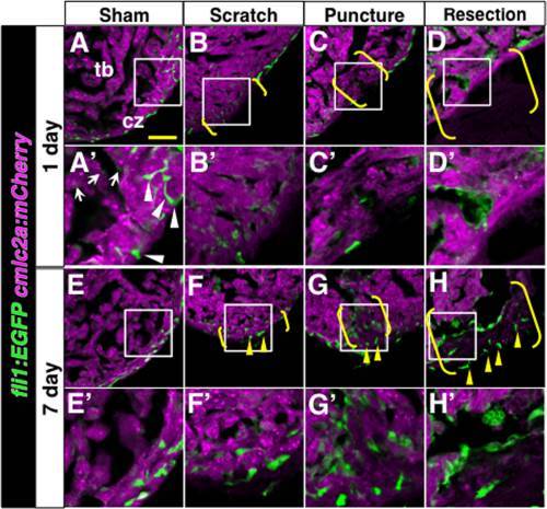
 Analysis of endothelial cells after different damages to the heart. The fli1:EGFP signal (green, endothelial cells) and the cmlc2a:mCherry signal (magenta, cardiomyocytes) at 1 day (A–D, A′–D′) and 7 days (E–H, E′–H′) after various injuries. A′–H′ show close-up images of boxed areas in A–H. White arrowheads and arrows in A′ point to thick and thinner fli1:EGFP signals, respectively. For simplicity, only A′ is labeled. Yellow arrowheads point to the fli1:EGFP signals at the surface of injury sites. Brackets indicate the injury sites. Scale bar = 50 µm. cz, compact zone; tb, trabecular zone. | 
                
                    
                        Expression Data


                        Expression Detail


                        Antibody Labeling


                        Phenotype Data


                        Phenotype Detail


                        Acknowledgments


This image is the copyrighted work of the attributed author or publisher, and
ZFIN has permission only to display this image to its users.
Additional permissions should be obtained from the applicable author or publisher of the image.
      Full text @ Dev. Dyn.


        