
Fig. 3
- ID
- ZDB-FIG-150122-22
- Publication
- Huang et al., 2013 - Nonmuscle myosin II-B (myh10) expression analysis during zebrafish embryonic development
- Other Figures
- All Figure Page
- Back to All Figure Page
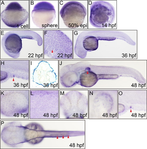
Whole mount in situ hybridization analysis of zebrafish embryos (1 cell-48 hpf) using antisense Danio rerio myh9a probe. (A) 1 cell, lateral view, strong staining. (B) Sphere, lateral view. (C) 50% epiboly, lateral view. (D) 14 hpf, lateral view, epidermis. (E) 22 hpf, lateral view, epidermis, enveloping layer (EVL). (F) 22 hpf, lateral view, caudal trunk, EVL (arrowhead). (G) 36 hpf, lateral view, epidermis of the whole body. (H) 36 hpf, lateral view, urogenital opening (arrowhead). (I) 36 hpf, transverse section of head, out layer staining. (J) 48 hpf, lateral view, pectoral fin (arrowhead). (K) 48 hpf, lateral view, eye epidermis. (L) 48 hpf, lateral view, trunk epidermis over the yolk tube elongation. (M) 48 hpf, ventral view, head epidermis. (N) 48 hpf, lateral view, pectoral fin epidermis. (O) 48 hpf, lateral view, urogenital opening (arrowhead). (P) 48 hpf, dorsal view, dorsal fin epidermis (arrowheads). |
| Gene: | |
|---|---|
| Fish: | |
| Anatomical Terms: | |
| Stage Range: | 1-cell to Long-pec |
Reprinted from Gene expression patterns : GEP, 13(7), Huang, Y., Wang, X., Wang, X., Xu, M., Liu, M., and Liu, D., Nonmuscle myosin II-B (myh10) expression analysis during zebrafish embryonic development, 265-270, Copyright (2013) with permission from Elsevier. Full text @ Gene Expr. Patterns