
Fig. 3
- ID
- ZDB-FIG-150120-21
- Publication
- Sheets et al., 2014 - Characterization of Ribeye Subunits in Zebrafish Hair Cells Reveals That Exogenous Ribeye B-Domain and CtBP1 Localize to the Basal Ends of Synaptic Ribbons
- Other Figures
- All Figure Page
- Back to All Figure Page
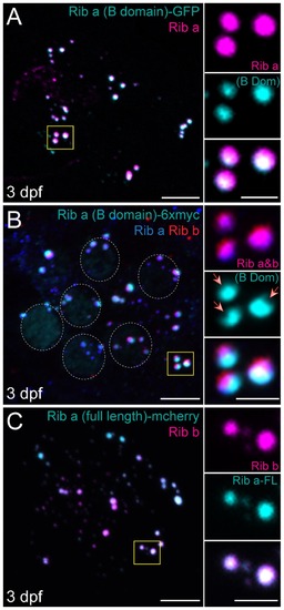
Ribeye B-domain/CtBP2s localizes to synaptic ribbons at 3 dpf. Representative images of immunolabel or fluorescent tag in posterior lateral line NM1 hair cells of 3 dpf larvae. Scale bars: 3 μm (main panels), 1 μm (insets). (A) Ribeye (B-domain)-GFP (cyan) and Ribeye a antibody labeling of synaptic ribbons (magenta). Note that B-domain-GFP does not completely colocalize with Ribeye a immunolabel. (B) Ribeye (B-domain)-myc (cyan), Ribeye a (blue), and Ribeye b (red) immunolabel. Dashed circles indicate nuclear localization of B-domain. Note that Ribeye a and b immunolabel colocalizes with each other (magenta), but only partially colocalizes with B-domain (white). Red arrows indicate resolvable indentations in the synaptic ribbons that do not contain exogenous B-Domain, but do contain Ribeye. (C) Ribeye a (full length)-mcherry (cyan) and Ribeye b immunolabel (magenta). Full length exogenous Ribeye a-mcherry appears throughout the synaptic ribbon and colocalizes with Ribeye b immunolabel (white). |
| Antibodies: | |
|---|---|
| Fish: | |
| Anatomical Term: | |
| Stage: | Protruding-mouth |