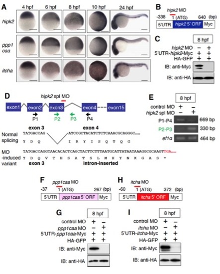
The expression patterns of zebrafish hipk2, ppp1caa, and itch and the effects of hipk2 MOs, ppp1caa MO, and itcha MO. Related to Figure 1.
(A) hipk2 mRNA, ppp1caa mRNA, and itcha mRNA are ubiquitously expressed throughout early embryogenesis. Panels show lateral views of whole mount in situ hybridization of hipk2, ppp1caa, and itcha in zebrafish embryos fixed at the indicated stages. Scale bar = 200 μm.
(B, C) hipk2 MO blocks translation of the hipk2 gene. To examine the effect of hipk2 MO, mRNA including the 5′UTR and 5′coding region (1–640 bp) of the hipk2 gene fused in-frame with a Myc-tag (B, 5′UTR-hipk2-Myc) was injected into zebrafish embryos with HA-GFP and hipk2 MO. The hipk2 MO annealing site is indicated by the red line in (B). In (C), embryo extracts were prepared at the indicated stages and then immunoblotted with anti-Myc and anti-HA antibodies.
(D, E) hipk2 spl MO inhibits the proper splicing of hipk2 mRNA. The genomic structure of the hipk2 gene is shown in (D). The hipk2 spl MO annealing site (red line), and hipk2 primers (P1: 5′- gaggtgctggagttcctgggtcga-3′, P2: 5′- ctctttatcccggagcatca-3′, P3: 5′- gcattcctgctcaataaggg-3′, and P4: 5′- ctctgctggcaagccctgcgtttg-3′) were used to monitor the effects on splicing by RT-PCR. Total RNA was extracted from 20 embryos at the indicated stages, reverse transcribed, and amplified by PCR. PCR of mRNA isolated from control MO-injected embryos using primers P1 and P4 produced a 669 bp band that corresponds to the correctly spliced transcript (E). Injection of hipk2 spl MO reduced the expression of this transcript. PCR of mRNA from embryos injected with hipk2 spl MO using primers P2 and P3 produced a 330 bp band (E). This product was mis-spliced (intron inserted) as shown in (D) and corresponds to a protein with a nonsense mutation immediately after exon 3. Elongation factor 1 alpha (ef1&alpjha;) mRNA was used as an internal control.
(F, G) ppp1caa MO blocks the translation of the ppp1caa gene in zebrafish embryos. To examine the effect of ppp1caa MO, mRNA including the 5′UTR and 5′coding region (1–267 bp) of the ppp1caa gene fused in-frame with a Myc-tag
(F, 5′UTR-ppp1caa-Myc) was injected into zebrafish embryos with HA-GFP and ppp1caa MO. The ppp1caa MO annealing site is indicated by the red line in (F). In (G), Embryo extracts were prepared at the indicated stages and then immunoblotted with anti-Myc and anti-HA antibodies.
(H, I) itcha MO blocks the translation of the itcha gene in zebrafish embryos. To examine the effect of itcha MO, mRNA including the 5′UTR and 5′coding region (1–372 bp) of the itcha gene fused in-frame with a Myc-tag (H, 5′UTR-itcha-Myc) was injected into zebrafish embryos with HA-GFP and itcha MO. The itcha MO annealing site is indicated by the red line in (H). In (I), embryo extracts were prepared at the indicated stages and then immunoblotted with anti-Myc and anti-HA antibodies.
|
