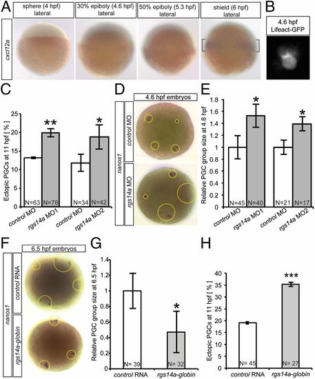
The role of Rgs14a in the temporal control of migration initiation. (A) Whole-mount in situ hybridization of embryos with a cxcl12a antisense RNA probe showing the initiation of transcription of the chemokine between 5.3 and 6.0 hpf. (B) Premigratory PGCs develop polarized actin brushes. (C) Morpholino-mediated knockdown of rgs14a leads to an increase in the average number of ectopic PGCs at 11 hpf (number of ectopic PGCs divided by total PGC number; Fig. S3). (D and E) PGC groups at 4.6 hpf (dotted circles) in Rgs14a knockdown embryos cover 53% larger areas than those in control embryos. (F and G) Early overexpression of Rgs14a, using the Xenopus globin32UTR, leads to significantly smaller PGC group areas at 6.5 hpf and an increase in the average number of ectopic PGCs at 11 hpf; target defined analogous to Fig. S3 (H). PGC groups in D–G were labeled with a nanos1 antisense RNA probe. Control embryos in F–H expressed a pa-(photoactivatable)-gfp-globin32UTR mRNA. For E and G, PGC group sizes were averaged for each embryo, and 32–45 embryos were included per experimental point. n = number of embryos analyzed. (C and H) *P < 0.05, **P < 0.01, and ***P < 0.001 using Student t test. (E and G) *P < 0.05 using Mann–Whitney u test. Error bars indicate SEM.
|
